Figure 2
Submitral Ventricular Pseudoaneurysm: Unusual and Late Complication of Cardiac Surgery
Marzia Cottini*, Amedeo Pergolini, Giordano Zampi, Vitaliano Buffa, Paolo Giuseppe Pino, Federico Ranocchi, Riccardo Gherli, De Marco Marina, Carlo Contento, Myriam Lo Presti and Francesco Musumeci
Published: 21 January, 2017 | Volume 1 - Issue 1 | Pages: 001-004

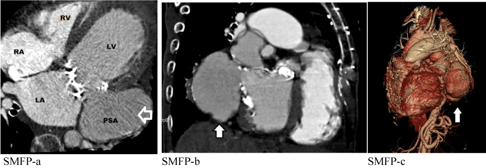
Figure 2:
2: a) Multi detector Computed Tomography, short-axis view showing sub mitral left ventricle pseudoaneurysm (arrow); b) long-axis view; c) multi detector computed tomography 3-D reconstruction of heart and SLVP (arrow).
Read Full Article HTML DOI: 10.29328/journal.hacr.1001001 Cite this Article Read Full Article PDF
More Images
Similar Articles
-
Submitral Ventricular Pseudoaneurysm: Unusual and Late Complication of Cardiac SurgeryMarzia Cottini*,Amedeo Pergolini,Giordano Zampi,Vitaliano Buffa,Paolo Giuseppe Pino,Federico Ranocchi,Riccardo Gherli,De Marco Marina,Carlo Contento,Myriam Lo Presti,Francesco Musumeci. Submitral Ventricular Pseudoaneurysm: Unusual and Late Complication of Cardiac Surgery . . 2017 doi: 10.29328/journal.hacr.1001001; 1: 001-004
Recently Viewed
-
‘Life-Changing Bubbles’ – How carbonated water can relieve swallowing problems for many dysphagia sufferers worldwideJohn Mirams. ‘Life-Changing Bubbles’ – How carbonated water can relieve swallowing problems for many dysphagia sufferers worldwide. J Addict Ther Res. 2023: doi: 10.29328/journal.jatr.1001024; 7: 001-004
-
Cognitive behavioral therapy treatment for drug addictionAlya Attiah Alghamdi*. Cognitive behavioral therapy treatment for drug addiction. J Addict Ther Res. 2023: doi: 10.29328/journal.jatr.1001025; 7: 005-007
-
Patient’s perception of the benefits of long-term opioids: Reinforcement associated with short-term effectsJames P Robinson*. Patient’s perception of the benefits of long-term opioids: Reinforcement associated with short-term effects. J Addict Ther Res. 2023: doi: 10.29328/journal.jatr.1001026; 7: 008-011
-
Internet Addiction and its Relationship with Attachment Styles Among Tunisian Medical StudentsRim Masmoudi*, Ahmed Mhalla, Amjed Ben Haouala, Wael Majdoub, Jawaher Masmoudi, Badii Amamou, Lotfi Gaha. Internet Addiction and its Relationship with Attachment Styles Among Tunisian Medical Students. J Addict Ther Res. 2023: doi: 10.29328/journal.jatr.1001027; 7: 012-018
-
Impact of Balanced Lifestyles on Childhood Development: A Study at CrècheP Vasundhara,P Nagaraju*. Impact of Balanced Lifestyles on Childhood Development: A Study at Crèche. J Addict Ther Res. 2024: doi: 10.29328/journal.jatr.1001028; 8: 001-008
Most Viewed
-
Feasibility study of magnetic sensing for detecting single-neuron action potentialsDenis Tonini,Kai Wu,Renata Saha,Jian-Ping Wang*. Feasibility study of magnetic sensing for detecting single-neuron action potentials. Ann Biomed Sci Eng. 2022 doi: 10.29328/journal.abse.1001018; 6: 019-029
-
Evaluation of In vitro and Ex vivo Models for Studying the Effectiveness of Vaginal Drug Systems in Controlling Microbe Infections: A Systematic ReviewMohammad Hossein Karami*, Majid Abdouss*, Mandana Karami. Evaluation of In vitro and Ex vivo Models for Studying the Effectiveness of Vaginal Drug Systems in Controlling Microbe Infections: A Systematic Review. Clin J Obstet Gynecol. 2023 doi: 10.29328/journal.cjog.1001151; 6: 201-215
-
Causal Link between Human Blood Metabolites and Asthma: An Investigation Using Mendelian RandomizationYong-Qing Zhu, Xiao-Yan Meng, Jing-Hua Yang*. Causal Link between Human Blood Metabolites and Asthma: An Investigation Using Mendelian Randomization. Arch Asthma Allergy Immunol. 2023 doi: 10.29328/journal.aaai.1001032; 7: 012-022
-
Impact of Latex Sensitization on Asthma and Rhinitis Progression: A Study at Abidjan-Cocody University Hospital - Côte d’Ivoire (Progression of Asthma and Rhinitis related to Latex Sensitization)Dasse Sery Romuald*, KL Siransy, N Koffi, RO Yeboah, EK Nguessan, HA Adou, VP Goran-Kouacou, AU Assi, JY Seri, S Moussa, D Oura, CL Memel, H Koya, E Atoukoula. Impact of Latex Sensitization on Asthma and Rhinitis Progression: A Study at Abidjan-Cocody University Hospital - Côte d’Ivoire (Progression of Asthma and Rhinitis related to Latex Sensitization). Arch Asthma Allergy Immunol. 2024 doi: 10.29328/journal.aaai.1001035; 8: 007-012
-
An algorithm to safely manage oral food challenge in an office-based setting for children with multiple food allergiesNathalie Cottel,Aïcha Dieme,Véronique Orcel,Yannick Chantran,Mélisande Bourgoin-Heck,Jocelyne Just. An algorithm to safely manage oral food challenge in an office-based setting for children with multiple food allergies. Arch Asthma Allergy Immunol. 2021 doi: 10.29328/journal.aaai.1001027; 5: 030-037

If you are already a member of our network and need to keep track of any developments regarding a question you have already submitted, click "take me to my Query."


