
More Information
Submitted: May 14, 2026 | Accepted: June 02, 2026 | Published: June 03, 2026
Citation: Mascarenhas O, Sarvesh A, Paunikar S. Evaluating a Novel Intranasal Drug Delivery System for Procedural Sedation in Adults Undergoing Endoscopic Procedures – A Prospective, Comparative Study. Int J Clin Anesth Res. 2026; 10(1): 1-3. Available from:
https://dx.doi.org/10.29328/journal.ijcar.1001036
DOI: 10.29328/journal.ijcar.1001036
Copyright license: © 2026 Mascarenhas O, et al. This is an open access article distributed under the Creative Commons Attribution License, which permits unrestricted use, distribution, and reproduction in any medium, provided the original work is properly cited.
Keywords: Massive hydrocephalus; Pediatric anesthesia; Magnetic Resonance Imaging (MRI); Pediatric MRI sedation; Difficult airway management; Raised intracranial pressure; Neuroanesthesia; Airway challenges; MRI compatible anesthesia; Pediatric neuroimaging

Anaesthesia for Magnetic Resonance Imaging in a Pediatric Patient with Massive Hydrocephalus
Oswald Mascarenhas, Abinav Sarvesh* and Saely Paunikar
Anaesthesiology, Lilavati Hospital and Research Center, Mumbai, Maharashtra, India
*Address for Correspondence: Abinav Sarvesh, Anaesthesiology, Lilavati Hospital and Research Center, Mumbai, Maharashtra, India, Email: [email protected]
Children and infants undergoing Magnetic Resonance imaging (MRI) often require anaesthesia or sedation to ensure immobility during the procedure. The concomitant presence of other comorbidities in this population poses additional risks during the procedure. We report the case of an 11-month-old male child with a body weight of 18 kgs with a head circumference of 88 cms posted for MRI brain. Anaesthetic considerations of massive hydrocephalus mainly include distorted anatomy, difficult airway, and raised Intracranial Pressure (ICP). We discuss the strategies available for similar scenarios and the rationale for avoiding sedation in this particular case.
Massive hydrocephalus is a challenging neurosurgical condition characterized by excessive accumulation of cerebrospinal fluid leading to progressive enlargement of the ventricular system and increased head circumference. Pediatric patients with severe hydrocephalus frequently present significant anesthetic challenges due to difficult airway management, positioning difficulties, and the risk of elevated intracranial pressure. Magnetic resonance imaging (MRI) is essential for diagnostic evaluation and surgical planning; however, maintaining immobility during MRI often necessitates sedation or general anesthesia. This case report describes the successful completion of brain MRI without sedation or general anesthesia in an infant with massive hydrocephalus and discusses important anesthetic considerations.
An eleven-month-old male child presented to our institute with progressive enlargement of the head and irritability. The child was delivered full term by cesarean section in view of hydrocephalus. Post-delivery, he was shifted to the neonatal intensive care unit and advised for further management, but later the patient was lost to follow-up. Currently, the child weighs 18 kgs, and the head circumference is 88 cms. He can lie in supine only, cries occasionally, and has sun-downing eyes. Neurological examination showed delayed milestones but no acute signs of cardio-respiratory compromise. MRI of the brain was planned to assess ventricular dilatation and rule out other causes before neurosurgical intervention. Due to the child’s massive head size, a difficult airway and raised ICP were anticipated, and hence we decided against general anaesthesia or sedation. Image 1.0 displays the massive head size of the child.
Before the procedure, we discussed with the parents regarding the current condition of the child and the risks of providing anaesthesia or sedation. So plan A was to do the procedure without anaesthesia, and plan B was to electively intubate the child in the operating theatre and then shift to MRI. An intravenous line was already placed in the ward. Proceeding with plan A, the MRI room was prepared with MRI-compatible monitors, emergency drugs loaded, machine and airway cart checked. The child and the mother were then taken inside the MRI room. The child was adequately swaddled and comforted with parental presence. Considering the massive head circumference, the child was not very active and was stable during the scans. The procedure took 40 minutes. Since the child moved twice during the scan, those two sequences were taken again. MRI was performed successfully without anesthesia, with good image quality obtained and no adverse events during the scan.
Children and infants undergoing Magnetic Resonance imaging (MRI) often require anaesthesia or sedation in order to ensure immobility, minimise motion artifacts, and maintain image quality during the procedure. The presence of other comorbidities and their manifestations in this population poses additional risks during the procedure. One such common condition encountered is hydrocephalus. These patients present a unique set of challenges mainly due to altered cranial anatomy, raised ICP, and potential airway difficulties [1]. So the anaesthesia technique must strike a balance between providing motion restriction for the procedure and avoiding the risks associated with sedation or general anaesthesia for the patients. Children with hydrocephalus commonly have distorted airway anatomy due to the massive head size, which causes limited neck movement, difficulty in positioning the patient supine, as well as achieving the alignment of the right axes during intubation. Raised ICP is seen in this set of patients, and it further rises on crying, coughing, and during airway instrumentation [2]. MRI room with the magnetic field restricts the use of standard anaesthetic equipment and urgent airway interventions once the scan has started. The ideal anaesthesia technique should ensure airway safety, minimal rise in ICP, and stable hemodynamics. Various techniques available for pediatric MRI are listed in Table 1.0.
| Table 1: Various techniques available for pediatric MRI. | ||||
| Techniques available for pediatric MRI [3-7] | ||||
| 1)No anaesthesia -Natural sleep [3] -Immobilization |
||||
| 2) Sedation [4] | Dose | Onset | Duration of action | Comments |
| Oral chloral hydrate | 50 - 75 mcg/kg | 20 - 30 minutes | 60 - 120 minutes | Can cause prolonged sedation [5] |
| Intranasal dexmedetomidine | 2 - 4 mcg/kg via atomizer | 20 - 25 minutes | 60 - 120 minutes | Minimal respiratory depression |
| Intravenous dexmedetomidine | 1 mcg/kg over 10 minutes then 0.3 - 0.7 mcg/kg maintenance | 5 - 10 minutes | 30 - 60 minutes | Bradycardia, Hypotension |
| Intravenous propofol | Bolus 1 - 2 mg/kg, then 25 - 75 mcg/kg/min | 30 - 60 seconds | 5 - 10 minutes | Respiratory and cardiovascular depression |
| Intravenous midazolam | 0.05 - 0.1 mg/kg | 2 - 3 minutes | 30 - 45 minutes | Respiratory depression |
| Intravenous ketamine | 0.5 - 2 mg/kg | 30 - 60 seconds | 10 - 20 minutes | Increased secretion and emergence delirium |
| 3)General anaesthesia -Endotracheal intubation -Laryngeal mask airway |
||||
Specific considerations for massive hydrocephalus start with the positioning of the patient. Careful padding and neutral head alignment are required to prevent venous obstruction and a rise in ICP. These patients are anticipated to have a difficult airway, so MRI-compatible airway equipment should be kept ready. MRI-compatible monitors and an anaesthesia machine should be used [6,7]. Control of ICP is essential for smooth induction and extubation. Avoidance of hypoxia, hypercarbia, and coughing is essential. It is also essential to maintain stable blood pressure and cerebral perfusion by avoiding hypotension and a rise in ICP. Conducting an MRI on the day of surgery can be beneficial, as an MRI can be conducted after endotracheal intubation, and later surgery can be done on the same day. This avoids the need for another intubation for a surgical procedure.
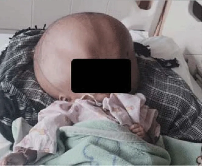
Figure 1: Massive hydrocephalus with increased head circumference.
Anaesthesia for pediatric MRI with massive hydrocephalus poses unique anaesthetic challenges. Sedation or general anesthesia is often necessary, but careful patient selection may allow successful imaging without pharmacological interventions, as in this case. An individual approach that balances the need for immobility against risks of airway compromise is crucial.
- Kumari N, Sai A, Sharma A, Kachhap A, Singh D, Kumar T. Anesthetic management in a case of huge hydrocephalus. Annals of African Medicine 2024; 23: 100–103. Available from:https://doi.org/10.4103/aam.aam_171_23
- Krovvidi H, Flint G, Williams AV. Perioperative management of hydrocephalus. BJA Education 2018; 18: 140–146. Available from: https://doi.org/10.1016/j.bjae.2018.01.007
- Harrington SG, Jaimes C, Weagle KM, Greer MC, Gee MS. Strategies to perform magnetic resonance imaging in infants and young children without sedation. Pediatric Radiology 2022; 52: 374–381. Available from:https://doi.org/10.1007/s00247-021-05062-3
- Mahmoud M, Mason KP. Dexmedetomidine: review, update, and future considerations of pediatric perioperative and periprocedural applications. Anesthesia & Analgesia 2015; 121: 470–482. Available from: https://doi.org/10.1093/bja/aev226
- American Society of Anesthesiologists. Practice guidelines for moderate procedural sedation and analgesia. Anesthesiology 2018; 128: 437–479. Available from: https://doi.org/10.1097/aln.0000000000002043
- Guimarães Ferreira Fonseca L, Garbin M, Bertolizio G. Anesthesia for pediatric magnetic resonance imaging: a review of practices and current pathways. Curr Opin Anaesthesiol. 2023 Aug 1;36(4):428-434.Available from: https://doi.org/10.1097/ACO.0000000000001267
- Ertan E, Eroğlu B. Anesthesia practices during magnetic resonance ımaging in pediatric patients. Surg Child. 2024;1(3):57-62