
More Information
Submitted: March 31, 2025 | Approved: April 21, 2025 | Published: April 23, 2025
How to cite this article: Warik A, Sarvesh A. Anesthesia Considerations in Scoliosis Surgery - A Clinical Communication. Int J Clin Anesth Res. 2025; 9(1): 023-025. Available from:
https://dx.doi.org/10.29328/journal.ijcar.1001030
DOI: 10.29328/journal.ijcar.1001030
Copyright License: © 2025 Warik A, et al. This is an open access article distributed under the Creative Commons Attribution License, which permits unrestricted use, distribution, and reproduction in any medium, provided the original work is properly cited.

Anesthesia Considerations in Scoliosis Surgery - A Clinical Communication
Anagha Warik and Abinav Sarvesh*
DNB, Anaesthesiology Resident, India
*Address for Correspondence: Abinav Sarvesh, DNB, Anaesthesiology Resident, India, Email: [email protected]
Scoliosis is a condition of abnormal lateral curvature of the spinal column greater than 10 degrees as measured by Cobb’s angle. The two main groups of scoliosis are idiopathic scoliosis and non-idiopathic scoliosis. The diagnosis of an idiopathic scoliosis is made if a non-idiopathic one has been excluded. Idiopathic adolescent scoliosis is a more common form. Neuromuscular scoliosis, which is a type of non-idiopathic scoliosis, is characterized by diverse muscular and neurological impairments. Anesthesia and surgery for neuromuscular scoliosis have a higher risk of perioperative complications than for idiopathic cases.
Scoliosis is a condition of abnormal lateral curvature of the spinal column greater than 10 degrees as measured by Cobb’s angle. Patients with severe scoliosis are at higher risk of respiratory failure, impaired ventilation-perfusion distribution, airway obstruction, which can lead to pulmonary dysfunction, especially in patients with adult idiopathic scoliosis [1].
The surgical correction of any scoliosis is indicated to prevent curve progression or the progression of restrictive lung disease and improve respiratory function [1]. Other indications include significant impact on the patient’s quality of life, usually due to poor sitting balance, back or Costo-pelvic pain, respiratory complications, or problems with self-care and feeding [2].
With this in mind, preoperative assessment, especially airway evaluation, is necessary for patients with scoliosis prior to administration of anesthesia.
Preoperative evaluation
Pulmonary Function Tests (PFTs) diagnose restrictive pulmonary deficits by demonstrating decreased Forced Expiratory Volume in one second (FEV1), decreased Forced Vital Capacity (FVC) with a normal FEV1/FVC ratio. Total Lung Capacity (TLC) is also decreased in patients with restrictive pulmonary disease [3] (Table 1).
| Table 1: Assessment of severity of restrictive lung disease using PFT [4]. | ||
| Degree of disease | % predicted FVC | % predicted TLC |
| Mild | 70%- lower limits of normal | 70%-lower limits of normal |
| Moderate | 60% - 69% | 60% - 69% |
| Moderately severe | 50% - 59% | < 50% |
| Severe | 34% - 49% | < 50% |
| Very severe | < 34% | < 50% |
| FVC: Forced Vital Capacity; TLC: Total Lung Capacity | ||
Because of the potential for significant pulmonary, cardiac, and neurological comorbidities associated with advanced scoliosis, a detailed past medical history and physical examination for patients undergoing scoliosis surgery is essential.
In spine evaluation, the lateral curvature is usually right-sided in the adolescent age group [5]. In case of left sided curvature is more common in the infantile age group [5]. There is an increased likelihood of other congenital comorbidities of the genitourinary, cardiac systems in cases of congenital scoliosis [6]. Measurement of Cobb’s angle is a key indicator in assessing spinal curvature, particularly in scoliosis, is important. Using advancements in automated and AI-based methods significantly improves the accuracy and efficiency of Cobb angle measurement for spinal curvature assessment [7].
A thorough pulmonary function evaluation is important, including incentive spirometry apart from chest x-ray and peripheral capillary oxygen saturation. Decreased lung volumes due to restriction from diaphragm and chest wall movement because of alteration of the thoracic cavity from scoliosis, leading to poor ventilation distribution, hypoxemia, and hypercarbia [8].
Most patients with mild or developing scoliosis can tolerate general anesthesia without complications if no cardiopulmonary abnormalities are found. Alternatively, in cases where there are no abnormalities present but scoliosis has manifested curvature in the trachea, the suggested course of action is nasotracheal intubation. Cardiovascular complications though less common, can be more severe than pulmonary complications. Due to compression of heart there is a reduction in stroke volume. Also, scoliosis is associated with many diseases of heart such as cardiomyopathy, congenital heart disease or valve diseases [6]. ECG and 2D echocardiogram should be done. A detailed neurological examination is essential due to medicolegal issues, in case of postoperative neurological deficit development. Severity of scoliosis is measured by Cobb’s angle on a standing X-ray of the thoracolumbar spine (Table 2).
| Table 2: Severity of scoliosis and presentation [9]. |
| Severity of scoliosis and presentation |
| < 10° - no symptoms > 25° - increase in pulmonary artery pressure > 40° - consider surgical intervention > 70° - significant decrease in lung volume > 100° - dyspnea on exertion > 120° - alveolar hypoventilation, chronic respiratory failure |
Anesthesia and intraoperative management
Anesthesia for scoliosis is very challenging for the anesthetic team due to its complications, such as prone positioning, big incision, high blood loss, Intraoperative Neurophysical Monitoring (IONM), long surgical times, fluid shift, and body temperature loss. Total Intravenous Anesthesia (TIVA), most often the combination of propofol and remifentanil, is the most suitable and safe method, especially in the case of IONM [10]. For this purpose, secure sufficient IV line or central venous catheterization can also be considered. During TIVA, depth of anesthesia is important to prevent overdosing. Non-depolarizing muscle relaxants can be administered safely. Rocuronium along with its antagonist Suggamadex, represents a safe combination to provide complete recovery post-operation and allow reliable IONM. The prone position is associated with several complications caused by raised intrathoracic or intraabdominal pressure. Postoperative visual loss is a rare but serious complication.
Patients undergoing scoliosis surgery are at a higher risk of extensive blood loss due to prolonged surgeries or bones with lower density. Tranexamic acid is one of the most widely used antifibrinolytic used for scoliosis surgery to reduce blood loss [11]. The recommended prophylactic dose is 15 mg/kg IV, and the maintenance dose is around 1-20 mg/kg/h [11]. Blood transfusion is started once the Hb reaches 7 gm/dl. Severe hypothermia due to long surgeries with a decrease in temperature of more than 2.5 °C from the baseline interferes with IONM. Other adverse effects include prolonged metabolism of anesthetic agents, coagulopathy with higher blood loss, and wound or respiratory infections [12] (Figure 1).
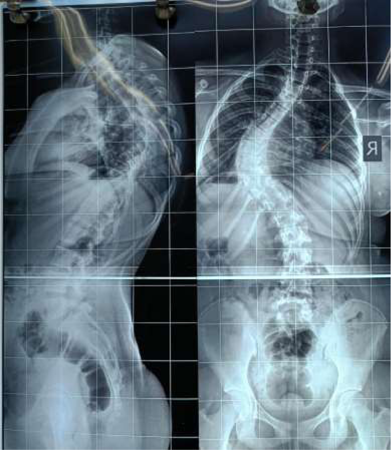
Figure 1: Lateral and AP X-ray of a scoliosis patient.
Intraoperative nerve monitoring
IONM is an important method of assessing neural structural and functional integrity. Specialized spinal cord function monitoring can be achieved by measuring evoked potentials. These are created by stimulating a peripheral nerve and measuring signals generated in the somatosensory cortex (somatosensory evoked potentials – SSEPs) or stimulating near the motor cortex and measuring signals at the target muscle (motor evoked potentials – MEPs). Anesthetic management, including oxygenation, ventilation, and massive blood loss, can influence the IONM reproducibility. The anesthetic team has to create the best environment for IONM. All muscle relaxants interfere with motor evoked potentials (MEP’s). They are administered during the phase of induction to the anesthesia to facilitate airway securing. SSEPs and MEPs are disrupted by inhalational agents at greater than 0.5 MAC and also by nitrous oxide [13] (Table 3).
| Table 3: Effects of Various Anaesthetic Agents on Neuromonitoring [14]. | ||
| Anesthetic drug | Effect on SSEP | Effect on MEP |
| Isoflurane | +++ | +++ |
| Sevoflurane | +++ | +++ |
| Nitrous oxide | ++ | ++++ |
| Barbiturates | +++ | +++ |
| Benzodiazepines | ++ | ++ |
| Propofol | ++ | ++ |
| Ketamine | +/- | +/- |
| Fantanyl / Remifentanyl | No effect | No effect |
Postoperative care
Majority patients require post-operative ICU admission. However, surgeries under four hours can sometimes be managed at post Anesthesia care unit. Patients with neuromuscular scoliosis often suffer from pre-existing respiratory diseases or restrictive lung disease which leads to a risk of postoperative respiratory failure [6]. Proper equipment for difficult airway management should be available at the ICU in case of emergent reintubation. Reintubation or prolonged weaning is associated with ventilator associated pneumonia or tracheal stenosis. Hence, early and successful weaning should be prioritized. Postoperatively, ECG, non-invasive blood pressure, and peripheral oxygen saturation should be monitored. In case of hemodynamically unstable patients, invasive blood pressure should be measured. Postoperative pain management after scoliosis surgery can be challenging due to the large skin incisions and multiple osteotomies. Optimizing pain control for patient satisfaction and prevention of respiratory complications from hypoventilation is an important consideration. A multimodal approach to analgesia using acetaminophen (paracetamol), NSAIDs, gabapentin, ketamine, opioids and other available analgesics may improve outcomes. Patient-controlled analgesia has been shown to enhance patient satisfaction and recovery [15].
Anesthesia management for scoliosis surgery requires careful pre-operative assessment, tailored Anesthetic techniques, and meticulous intraoperative monitoring to ensure patient safety and facilitate successful surgery, including neurophysiological monitoring and blood conservation strategies.
- Shapiro F, Korman J, McPherson E. Does posterior scoliosis correction improve respiratory function in adolescent idiopathic scoliosis? A systematic review and meta-analysis. Eur Spine J. 2019;28(6):1295-1303.
- Loughenbury PR, Tsirikos A. Current concepts in the treatment of neuromuscular scoliosis: Clinical assessment, treatment options, and surgical outcomes. J Bone Joint Surg Am. 2018;100(22):e156.
- Vazquez-Nieves R, Fonseca-Ferrer V, Irizarry-Nieves J. Measuring restrictive lung disease severity using FEV1 vs TLC. Chest. 2023;163(1):e1-e9.
- Kynes JM, Evans FM, Hodgetts V, Wilson K. Surgical correction of scoliosis: Anesthetic considerations. Anesth Tutor Week. 2019;19(6):1-10.
- Konieczny MR, Senyurt H, Krauspe R. Epidemiology of adolescent idiopathic scoliosis. J Child Orthop. 2013;7(1):3-9. Available from: https://doi.org/10.1007/s11832-012-0457-4
- Furdock R, Brouillet K, Luhmann SJ. Organ system anomalies associated with congenital scoliosis: A retrospective study of 305 patients. J Pediatr Orthop. 2018;38(9):e563-e568.
- Chen J, Wang S, Yang G, Li E, Liang Z. A review of the methods on Cobb angle measurements for spinal curvature. Sensors (Basel). 2022;22(9):3258. Available from: https://www.mdpi.com/1424-8220/22/9/3258
- Lin Y, Tan H, Rong T, et al. Impact of thoracic cage dimension and geometry on cardiopulmonary function in patients with congenital scoliosis: A prospective study. Spine J. 2019;19(9):1486-1494.
- Hudec J, Prokopova T, Kosinova M, Gal R. Anesthesia and perioperative management for surgical correction of neuromuscular scoliosis in children: A narrative review. Paediatr Anaesth. 2023;33(1):5-13.
- Nguyen A, Mandavalli A, Diaz MJ, et al. Neurosurgical anesthesia: Optimizing outcomes with agent selection. J Neurosurg Anesthesiol. 2022;34(4):345–352.
- Alajmi T, Saeed H, Alfaryan K, Alakeel A, Alfaryan T. Efficacy of tranexamic acid in reducing blood loss and blood transfusion in idiopathic scoliosis: A systematic review and meta-analysis. J Spine Surg. 2017;3(4):531–540. Available from: https://pmc.ncbi.nlm.nih.gov/articles/PMC5760407/
- Safari A, Parsaei H, Zamani A, Pourabbas B. A semi-automatic algorithm for estimating Cobb angle. J Biomed Phys Eng. 2019;9(3):317–326. Available from: https://doi.org/10.31661/jbpe.v9i3jun.730
- Pajewski TN, Arlet V, Phillips LH. Current approach on spinal cord monitoring: The point of view of the neurologist, the anesthesiologist, and the spine surgeon. Spine J. 2016;16(5):e77–e85. Available from: https://pmc.ncbi.nlm.nih.gov/articles/PMC2072895/
- Soghomonyan S, Moran KR, Sandhu GS, Bergese SD. Anesthesia and evoked responses in neurosurgery. Front Pharmacol. 2014 Apr 14;5:74. Available from: https://doi.org/10.3389/fphar.2014.00074
- Wishart BD, Kivlehan E. Neuromuscular scoliosis: When, who, why and outcomes. Phys Med Rehabil Clin N Am. 2021;32:547–556. Available from: https://doi.org/10.1016/j.pmr.2021.02.007